Mohs Micrographic Surgery
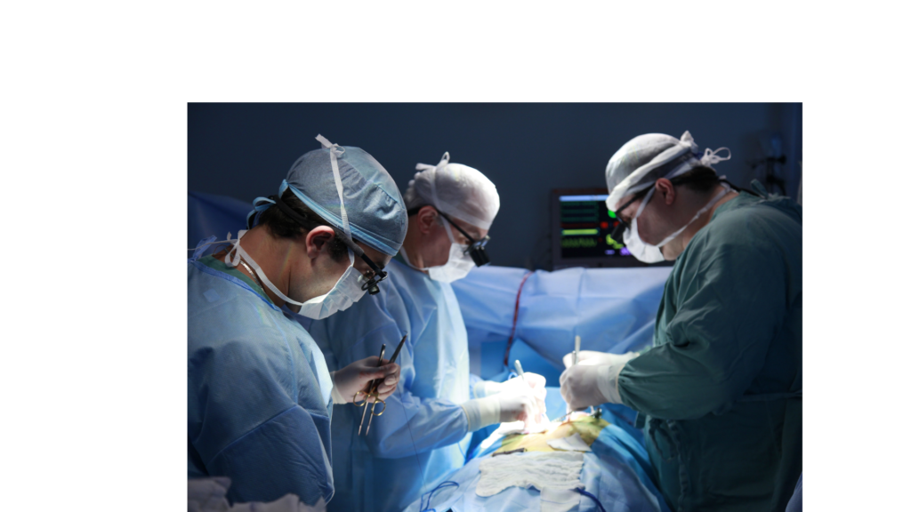
Mohs micrographic surgery is a technique for the removal of complex or ill-defined skin cancer with histologic examination of 100% of the surgical margins. It requires the integration of an individual functioning in two separate and distinct capacities: surgeon and pathologist. If either of these responsibilities is delegated to another physician or other qualified healthcare professional who reports the services separately, these codes should not be reported. The Mohs surgeon removes the tumor tissue and maps and divides the tumor specimen into pieces, and each piece is embedded into an individual tissue block for histopathologic examination. Thus a tissue block in Mohs surgery is defined as an individual tissue piece embedded in a mounting medium for sectioning.
Mohs micrographic surgery is a highly specialized and precise technique commonly employed in dermatology for the treatment of certain types of skin cancers, particularly basal cell carcinoma, and squamous cell carcinoma. This surgical procedure is named after Dr. Frederic E. Mohs, who developed the technique in the 1930s. The primary goal of Mohs surgery is to remove the cancerous cells while sparing as much healthy tissue as possible.
Here’s an overview of how Mohs micrographic surgery is typically performed in a medical setting:
- Local Anesthesia: The patient is first administered local anesthesia to numb the area where the skin cancer is located. This ensures that the patient remains comfortable throughout the procedure.
- Tumor Removal: The surgeon then removes the visible tumor along with a very thin layer of surrounding tissue. The removed tissue is carefully marked and mapped for precise orientation.
- Tissue Processing: The excised tissue is divided into sections and processed in a way that allows the surgeon to examine the entire surgical margin. This often involves freezing the tissue and cutting it into thin horizontal sections.
- Microscopic Examination: Each section is examined under a microscope to check for the presence of cancer cells at the edges. If cancer cells are identified, their location is precisely marked on the map.
- Additional Layers, if Necessary: Based on the microscopic findings, the surgeon returns to the patient and selectively removes additional tissue from the areas where cancer cells were identified. This process is repeated until no cancer cells are detected at the margins.
- Wound Closure: Once the surgeon is confident that all cancer cells have been removed, the wound is closed. Depending on the size and location of the wound, this may involve natural healing, stitches, or more complex reconstructive procedures.
The main advantage of Mohs surgery is its high cure rate, often exceeding 99% for certain types of skin cancer. Additionally, it allows for the preservation of healthy tissue, which is especially critical in cosmetically sensitive areas such as the face.
Mohs surgery is commonly recommended for skin cancers with ill-defined borders, aggressive growth patterns, or those located in anatomically sensitive areas where tissue preservation is crucial. However, it may not be the preferred method for all skin cancer cases, and the decision to use Mohs surgery is based on factors such as the type of cancer, its location, and the individual patient’s health.
If repair is performed, use separate repair, flap, or graft codes. If a biopsy of suspected skin cancer is performed on the same day as Mohs surgery because there was no prior pathology confirmation of a diagnosis, then report a diagnostic skin biopsy (11102, 11104, 11106) and frozen section pathology (88331) with modifier 59 to distinguish from the subsequent definitive surgical procedure of Mohs surgery.
(If additional special pathology procedures, stains, or immunostains are required, see 88311-88314, 88342)
(Do not report 88314 in conjunction with 17311-17315 for routinely frozen section stain (eg, hematoxylin and eosin, toluidine blue) performed during Mohs surgery. When a nonroutine histochemical stain on frozen tissue is utilized, report 88314 with modifier 59)
(Do not report 88302-88309 on the same specimen as part of the Mohs surgery)
CPT Code: 17311 Mohs micrographic technique, including removal of all gross tumors, surgical excision of tissue specimens, mapping, color coding of specimens, microscopic examination of specimens by the surgeon, and histopathologic preparation including routine stain(s) (eg, hematoxylin and eosin, toluidine blue), head, neck, hands, feet, genitalia, or any location with surgery directly involving muscle, cartilage, bone, tendon, major nerves, or vessels; first stage, up to 5 tissue blocks.
CPT code 17311 is specifically used to describe the first stage of the Mohs micrographic technique when it involves the head, neck, hands, feet, genitalia, or any location with surgery directly involving muscle, cartilage, bone, tendon, major nerves, or vessels. Below is a more detailed breakdown of the coding guidelines along with an example:
CPT Code: 17311
- Description: Mohs micrographic technique, including removal of all gross tumors, surgical excision of tissue specimens, mapping, color coding of specimens, microscopic examination of specimens by the surgeon, and histopathologic preparation including routine stain(s) (e.g., hematoxylin and eosin, toluidine blue), head, neck, hands, feet, genitalia, or any location with surgery directly involving muscle, cartilage, bone, tendon, major nerves, or vessels; first stage, up to 5 tissue blocks.
Coding Guideline:
- Procedure Components:
- Removal of all gross tumors.
- Surgical excision of tissue specimens.
- Mapping and color coding of specimens.
- Microscopic examination of specimens by the surgeon.
- Histopathologic preparation, including routine stain(s) such as hematoxylin and eosin or toluidine blue.
- Anatomical Locations:
- The procedure is applicable to specific anatomical locations, including the head, neck, hands, feet, genitalia, or any location with surgery directly involving muscle, cartilage, bone, tendon, major nerves, or vessels.
- Stage of Procedure:
- This code specifically represents the first stage of the Mohs micrographic technique.
- Number of Tissue Blocks:
- Up to 5 tissue blocks are included in this code.
Example:
- A patient presents with a basal cell carcinoma on the forehead (head location). The Mohs micrographic technique is employed to remove the tumor.
- The surgeon removes the gross tumor along with a thin margin.
- Surgical excision of tissue specimens is performed.
- The excised tissue is mapped and color-coded for identification.
- The surgeon microscopically examines the tissue specimens in real time during the procedure.
- Histopathologic preparation, including routine staining (e.g., hematoxylin and eosin), is done to assess the tissue.
This entire process represents the first stage of the Mohs micrographic technique for this patient, and if the total number of tissue blocks does not exceed 5, CPT code 17311 will be used for billing purposes.
CPT Code ✚ 17312 each additional stage after the first stage, up to 5 tissue blocks (List separately in addition to code for primary procedure)
(Use 17312 in conjunction with 17311)
17313 Mohs micrographic technique, including removal of all gross tumors, surgical excision of tissue specimens, mapping, color coding of specimens, microscopic examination of specimens by the surgeon, and histopathologic preparation including routine stain(s) (eg, hematoxylin and eosin, toluidine blue), of the trunk, arms, or legs; first stage, up to 5 tissue blocks.
CPT Code: 17313
- Description: Mohs micrographic technique, including removal of all gross tumors, surgical excision of tissue specimens, mapping, color coding of specimens, microscopic examination of specimens by the surgeon, and histopathologic preparation including routine stain(s) (e.g., hematoxylin and eosin, toluidine blue), of the trunk, arms, or legs; first stage, up to 5 tissue blocks.
Coding Guideline:
- Procedure Components:
- Removal of all gross tumors.
- Surgical excision of tissue specimens.
- Mapping and color coding of specimens.
- Microscopic examination of specimens by the surgeon.
- Histopathologic preparation, including routine stain(s) such as hematoxylin and eosin or toluidine blue.
- Anatomical Locations:
- The procedure is applicable to specific anatomical locations, including the trunk, arms, or legs.
- Stage of Procedure:
- This code specifically represents the first stage of the Mohs micrographic technique.
- Number of Tissue Blocks:
- Up to 5 tissue blocks are included in this code.
Example:
- A patient has a squamous cell carcinoma on the arm. The Mohs micrographic technique is chosen as the treatment approach.
- The surgeon removes the gross tumor along with a thin margin.
- Surgical excision of tissue specimens is performed.
- The excised tissue is mapped and color-coded for identification.
- The surgeon microscopically examines the tissue specimens in real time during the procedure.
- Histopathologic preparation, including routine staining (e.g., hematoxylin and eosin), is done to assess the tissue.
This entire process represents the first stage of the Mohs micrographic technique for this patient, and if the total number of tissue blocks does not exceed 5, CPT code 17313 will be used for billing purposes.
CPT Code ✚ 17314 each additional stage after the first stage, up to 5 tissue blocks (List separately in addition to code for primary procedure)
(Use 17314 in conjunction with 17313).
CPT Code ✚ 17315 Mohs micrographic technique, including removal of all gross tumors, surgical excision of tissue specimens, mapping, color coding of specimens, microscopic examination of specimens by the surgeon, and histopathologic preparation including routine stain(s)
(eg, hematoxylin and eosin, toluidine blue), each additional block after the first 5 tissue blocks, any stage (List separately in addition to code for primary procedure) (Use 17315 in conjunction with 17311-17314)
Thank you for Visiting to Health Coding Hub
For any queries, please reach out to us at info@healthcodinghub.com
-
Previous Post
Thanksgiving Day ICD 10 CM Codes
-
Next Post
CPT 2024 Updates


